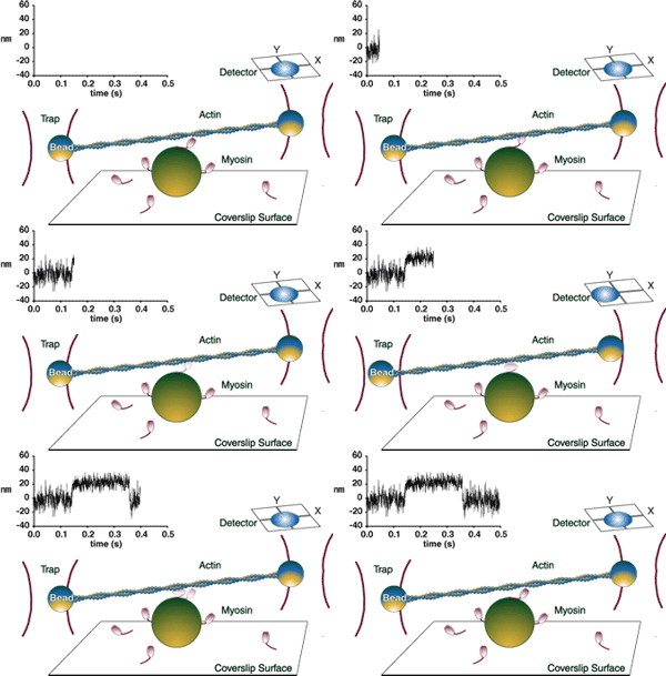
 The laser trap has the capacity to capture microscopic particles, such as 1 µm polystyrene beads, in solution. A trap is created by focusing a laser through a high numerical aperture objective, forming a trap at the focal point of the objective. This technique can be used to measure the molecular forces (piconewtons) and displacements (nanometers) generated by a single myosin molecular motor as it interacts with a single actin filament. For these measurements, two traps are formed by a computer-controlled acousto-optic deflector that timeshares the laser between two positions at 10 kHz. Then, a single NEM-modified myosin-coated bead is captured in each trap. With the ability to manipulate the position of the traps through software (developed by Dr. Joe Patlak), the investigator attaches a bead to each end of an actin filament. The actin filament is then lowered towards the motility surface where larger beads serve as a myosin platform. As the myosin pulls on the actin filament, the displacement of the actin filament is detected through the motion of one or both of the beads, the image of which is projected onto a quadrant photodiode detector (Guilford et al., 1997, Dupuis et al., 1997). Recently, we have developed a computer-based force clamp feedback system allowing us to characterize the effect of load on the mechanics and kinetics of a single myosin molecule. The laser trap has the capacity to capture microscopic particles, such as 1 µm polystyrene beads, in solution. A trap is created by focusing a laser through a high numerical aperture objective, forming a trap at the focal point of the objective. This technique can be used to measure the molecular forces (piconewtons) and displacements (nanometers) generated by a single myosin molecular motor as it interacts with a single actin filament. For these measurements, two traps are formed by a computer-controlled acousto-optic deflector that timeshares the laser between two positions at 10 kHz. Then, a single NEM-modified myosin-coated bead is captured in each trap. With the ability to manipulate the position of the traps through software (developed by Dr. Joe Patlak), the investigator attaches a bead to each end of an actin filament. The actin filament is then lowered towards the motility surface where larger beads serve as a myosin platform. As the myosin pulls on the actin filament, the displacement of the actin filament is detected through the motion of one or both of the beads, the image of which is projected onto a quadrant photodiode detector (Guilford et al., 1997, Dupuis et al., 1997). Recently, we have developed a computer-based force clamp feedback system allowing us to characterize the effect of load on the mechanics and kinetics of a single myosin molecule. |
Warshaw Laboratory – University of Vermont
